Metabolomic profiling using LC-Q-Orbitrap HRMS
In general, metabolomic studies on Salvia species in negative ionization mode tend to be more efficient than these in positive mode15. In this study, MS data analysis included the use of online and local databases provided by Compound Discoverer 2.1 software. Additionally, data collected from previous metabolomic studies on Salvia species were collected and merged into a mass list to be implemented as a local database. A total of 2704 substance peaks had been detected in S. fruticosa extracts in negative ion mode analysis. After filtering out the minor signals (Area < 104), there were 98 metabolites of which 95 were tentatively identified as shown in Supplementary Table S1.
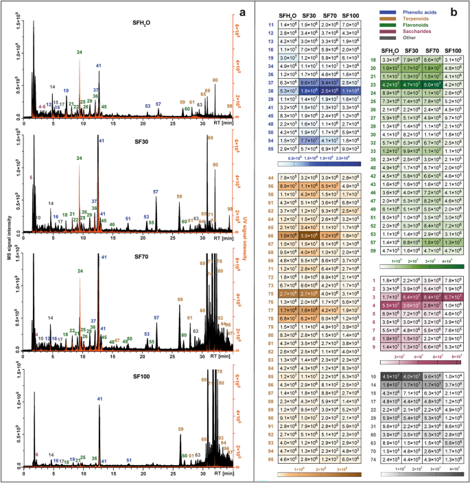
Metabolite profiles of each extract were juxtaposed and presented in Fig. 1a. The dominant groups changed among different extracts. In those obtained with solvents containing mostly water (SFH2O and SF30) the most abundant compounds were phenolic acids. The extraction efficiency of terpenoid compounds was consistent with increase of ethanol in used solvent owing to the non-polar nature of these compounds. Additionally, a heat map with the signal intensity of individual phytochemicals detected in four different S. fruticosa extracts is presented in Fig. 1b. The most numerous class of compounds detected in studied extracts was terpenoids with 35 compounds, followed by flavonoids (24 compounds), phenolic acids and derivatives (19 compounds), saccharides (9 compounds) and other such as fatty acids, carboxylic acids and unidentified compounds.
Total ion chromatograms obtained by LC-Q-Orbitrap in negative mode (black) combined with chromatograms registered by UV–Vis detector at 270 nm (orange) (a), set with heat map representing the mean MS peak area value of the identified compounds in four different S. fruticosa extracts: SFH2O–water extract; SF30–30% ethanol extract; SF70–70% ethanol extract; SF100–ethanol extract (b). For the identity of peaks, see Supplementary Table S1.
In the case of two most polar extracts (SFH2O and SF30) the phenolic acids were the most abundant classes in total peak area. This class was represented mainly by caffeic acid derivatives. The retention time (RT) of compound 16 with precursor ion [M-H]¯ at m/z 179.03419 was in line with the RT of the caffeic acid standard. It also generated characteristic major fragment at m/z 135.04414, due to loss of carbon dioxide. The deprotonated form of caffeic acid was detected in compounds 40 and 41, which were identified as sagerinic acid ([M-H]¯ at m/z 719.16210) and rosmarinic acid ([M-H]¯ at m/z 359.0773). Rosmarinic acid identification was additionally confirmed by comparison with the standard. The same ion or its loss had been observed for compounds 20, 37, 39, 44, 53, 57 and 58, which supported by comparison with the literature and MS2 fragmentation, were identified as salviaflaside ([M-H]¯ m/z 521.13012), salvianolic acid B ([M-H]¯ at m/z 717.14661), isosalvianolic acid B ([M-H]¯ at m/z 717.14667), salvianolic acid K ([M-H]¯ at m/z 555.11469), two salvianolic acid F isomers ([M-H]¯ at m/z 313.07205) and salvianolic acid C ([M-H]¯ at m/z 491.09863).
In the case of the two most non-polar extracts (SF70, SF100) the contribution of terpenoids was the highest, while in the remaining extracts this class accounted for about a quarter of the sum of the peak area of the identified compounds. This class was represented mainly by diterpenoids, which were the most varied non-polar class of compounds identified in studied extracts. They were mostly abietane-type diterpenoids, for which fragmentation through negative ionization oftentimes included the removal of CO2 (-44 Da), CO (-28 Da), H2O (-18 Da), ·CH3 (15 Da). Compounds 59 ([M-H]¯ at m/z 345.17075) and 64 ([M-H]¯ at m/z 345.17100) both displayed ions attributable to the loss of carbon dioxide molecule (m/z 301.18097) and water molecule (m/z 283.17038 and m/z 283.17041), and were identified as rosmanol and epiisorosmanol. Compound 69 ([M-H]¯ at m/z 329.17580) was identified as carnosol based on its typical fragmentation pattern, starting with the loss of carbon dioxide (m/z 285.16604)12,15 and followed by the elimination of a methyl radical (m/z 270.16211). The same fragmentation pattern occurred in compound 80, identified as 12-metoxy carnosic acid ([M-H]¯ at m/z 345.20721) with fragments of 301.21689 and 286.19385. Compound 78, with a pseudomolecular ion at m/z 331.19153 [M-H]¯ was identified as carnosic acid owing to the presence of fragments corresponding to the loss of carbon dioxide and subsequent loss of an isopropyl radical (m/z 287.20175 and 244.14687). Compound 70 showed a precursor ion at [M-H]¯ at m/z 343.15524, which generated characteristic fragments m/z 315.16028 and m/z 299.160504 via the loss of ethylene and carbon dioxide, respectively. That allows us to identify compound 70 as rosmadial. Two pentacyclic triterpenoids were also detected in the tested extracts: compound 96 and 97, which were tentatively identified as betulinic acid and ursolic acid, respectively, with quasimolecular ions at ([M-H]¯ at m/z 455.35340). The presence of these triterpenoids was also reported in S. fruticosa by Jash et al.16.
In the extracts studied, especially those with a high water content (SFH2O, SF30), a significant share of oligosaccharides and sugar acids in the total peak area of the identified compounds was also noted. Compounds 1, 2 and 3 were identified tentatively as stachyose, raffinose and sucrose, as they are often major transport sugars in the Salvia species31. Compounds 4–8 were classified as sugar acids. The fragmentation pattern of compound 6 ([M-H]¯ at m/z 135.02875) was identical to that of l-threonic acid. Compound 8 ([M-H]¯ at m/z 149.0081) generated fragments m/z 72.99171, 59.01249 and 87.00734 that can be observed in negative ionization mode for l-( +)-tartaric acid.
Another major class of phytochemicals detected in S. fruticosa extracts was flavonoids. Most of the identified compounds belonging to this class have been assigned to flavones. Compound 24 was unambiguously identified as scutellarin by comparing the retention times, UV spectra and MS/MS fragmentation patterns with those of the commercial standard. Compounds 46 and 55 showed nearly the same precursor ions [M-H]¯ at m/z 299.0563 and 299.0562. Compound 46 produced most abundant fragments at m/z 284.03253 and 136.98682, similarly compound 55. These data correspond with the fragmentation pattern of hispidulin or diosmetin. Since there was a difference in retention time, both compounds could be present in S. fruticosa extracts. Compound 49 showed a precursor ion at [M-H]¯ at m/z 285.04065 that formed specific product ions at m/z 133.02834, 151.00261, 175.03903, in line with these reported for luteolin by Velamuri et al.17. Compound 52 ([M-H]¯ at m/z 327.21786) was identified as salvigenin (pectolinarigenin-7-methyl ether) as this flavone has been reported previously in S. fruticosa. Compound 54 yielded the base peak [M-H]¯ at m/z 269.04578. Precursor ion and product ions at m/z 117.03332 and 151.00264 confirmed that this compound is apigenin. Compound 56 gave the precursor ion [M-H]¯ at m/z 329.0668, indicating that its molecular formula was C17H14O7. It produced prominent fragment ions at m/z 299.01981 attributable to the loss of two methyl groups, and 271.02472, owing to the further elimination of carbon monoxide. Therefore, this peak was identified as jaceosidin. Compound 60 was identified as cisimaritin based on a precursor ion [M-H]¯ at m/z 313.07190 and the diagnostic product ions at m/z 298.04694 and 283.02478, indicating the loss of two methyl radicals and 255.02974 from the elimination of carbon monoxide. Compound 62 ([M-H]¯ at m/z 283.06137) corresponds to an apigenin derivate considering the fragment at m/z 268.03772 and 117.03318. Characteristic fragment ion at m/z 240.04193 formed by the loss of carbon monoxide led to compound 62 being identified as genkwanin. Fragmentation patterns of apigenin, hispidulin, cirsimaritin and genkwanin were consistent with those reported by Koutsoulas et al.12. Compound 50 was the only type of flavonol aglycon detected in studied extracts. With the precursor ion [M-H]¯ at m/z 315.0513 and main MS/MS fragment at m/z 300.02756 resulting from the loss of methyl group this compound was identified as isorhamnetin. Compound 30 with a pseudomolecular ion [M-H]¯ at m/z 609.18329 did not show any fragmentation, but since it was previously reported in S. fruticosa18,19, it was tentatively identified as flavanone—hesperidin. Flavonoid glycosides found in this study were mainly glucosides with characteristic fragment of 162 Da, glucuronides (176 Da) and rutinosides (308 Da). Luteolin glucoside (compound 22 with [M-H]¯ at m/z 447.09344) is present in most publications concerning the chemical composition of S. fruticosa extracts12,18,19,20,21. Compound 26 showed a precursor ion [M-H]¯ at m/z 491.0836 and was identified as isorhamnetin glucuronide, reported earlier in S. fruticosa only by Gürbüz et al.22. Compound 27 ([M-H]¯ at m/z 577.15668), identified as apigenin-rutinoside was also found in Greek sage by Cvetkovikj et al.21.
The presence of fatty acids was also observed in S. fruticosa extracts. Compounds 63 and 73 were identified tentatively as two polyunsaturated fatty acids. Compound 63 was assigned as dihydroxyoctadecadienoic acid (C18H31O4¯). Compound 73 produced precursor ion [M-H]¯ at m/z 295.22803 and characteristic fragments at m/z 277.21738 ([M-H-H2O]− and 195.13837 [M-(CHO-(CH2)4-CH3)-H]¯, indicating the position of the hydroxyl group at 13 carbon atom. Thus, it was identified as 13-hydroxy-9,11-octadecadienoic acid. Also, in S. fruticosa extracts the presence of the glucoside of tuberonic acid (m/z 387.16644) (compound 14) which is a growth hormone was observed.
Quantitative analysis of major phytochemicals
A quantification of the main phenolic compounds in various extracts of S. fruticosa of the dry weight of plant material (mg/g DW) is presented in Table 1. The content of caffeic acid, scutellarin, salvianolic acid B, rosmarinic acid, carnosic acid and carnosol was calculated based on calibration curves of authentic standards, while the content of other compounds was estimated in relation to the most similar available standard.
Overall, the most abundant compound in sage extracts is rosmarinic acid, which is a phenolic acid and a dimer of caffeic acid. The highest concentration of rosmarinic acid among all tested samples was found in SF70 (31.56 ± 1.88 mg/g DW), which is like the concentration of rosmarinic acid in S. fruticosa collected from Croatia (29.10 ± 0.21 mg/g DW), reported by Mervić et al.23. Even higher content (60.73 mg/g DW) was reported in methanolic extract from Greek variety of sage studied by Sarrou et al.19, but in this study the use of pure alcohol as a solvent did not result in the highest yield of rosmarinic acid. Rosmarinic acid concentration in the infusion (SFH2O) was much lower (4.96 ± 0.65 mg/g DW) than in other extracts which is not consistent with the findings of a similar comparison made for the Turkish variety of S. fruticosa by Tekin18. Caffeic acid was also detected in all studied extracts at similar concentrations (0.13–0.15 mg/g DW), which were ten times lower than those reported by Mervić et al.23. However, few salvianolic acids, which belong to major caffeic acid-derived trimers in sage plants, were present in greater amounts. The highest concentration of salvianolic acid B was obtained in SF70 and SF30 extracts (6.86 ± 0.93 mg/g DW and 6.52 ± 0.48 mg/g DW). Salvianolic acid K was the most abundant in ST30 extract with a concentration of 6.25 ± 1.0 mg/g DW. According to data presented by Cvetkovikj et al.21, the maximum concentration of salvianolic acid K among several studied Greek populations of S. fruticosa was 7.20 mg/g DW.
The most abundant terpenoid compounds in S. fruticosa are carnosic acid and carnosol, which both belong to the family of abietane diterpenoids12. The highest content of carnosic acid was observed in SF100 (14.82 ± 1.66 mg/g DW), followed by SF70 (13.88 ± 2.52 mg/g DW) which is not statistically different. These results are in line with the content measured in methanolic extract by Kallimanis et al.24 which was 12.5 ± 1.6 mg/g DW. The amount of carnosol in SF70 extract was 7.88 ± 1.33 mg/g DW and it was consistent with this reported by Sarrou et al.19. Salviol, the third most abundant terpenoid in studied extracts, is a meroterpenoid derived from abietane diterpenoid—ferruginol and is common in other Greek species of sage, e.g. S. pomifera25. This compound has not yet been reported in S. fruticosa; however it was present in most of studied extracts with the highest content: 7.37 ± 0.71 mg/g DW in SF70.
The third group of bioactives detected in S. fruticosa extracts were flavonoids. Scutellarin is one of the common flavonoids found in sage26. It was the most abundant flavonoid in the studied extracts with a similar yield: 7.77 ± 0.48 mg/g DW, 8.92 ± 1.56 mg/g DW and 7.35 ± 0.9 mg/g DW in SFH2O, SF30 and SF70 extracts, respectively. The concentrations of luteolin rutinoside and luteolin glucoside were similar in all studied extracts and ranged from 1.03 to 1.98 mg/g DW, which is not in line with data reported by Tekin et al.18, where the concentrations of these compounds in sage infusions were two to three times higher than in ethanol extracts.
Phenolic acids, flavonoids and terpenoids are typical bioactive compounds in S. fruticosa. As shown in Table 1, the extraction yield was highly affected by ethanol content in the solvent. The extraction with 70% ethanol provided the highest total yield of bioactives, in contrast to extraction with only water. The difference is clearly visible in the yield of phenolic acids and terpenoids, which in SF70 was three times higher and seven times higher, respectively. The maximum extraction yield of flavonoids was obtained with 30% ethanol, but it was only slightly higher than that obtained with 70% ethanol. Considering all groups of studied bioactives, 70% ethanol is concluded to be the best solvent among those tested for extraction of bioactive compounds from S. fruticosa.
Antioxidant activity
The presence of compounds exhibiting antioxidant activity in the plant material has become an important aspect defining its health-promoting properties. In the case of various species of sage, their high antioxidant activity is caused mainly by phenolic compounds. In the presented studies, the total antioxidant activity was determined for S. fruticosa extracts prepared with extractants of different polarity. In addition, the antioxidant activity was determined for selected phenolic compounds typical for sage and belonging to various classes of secondary metabolites such as phenolic acids, flavones and diterpenoids.
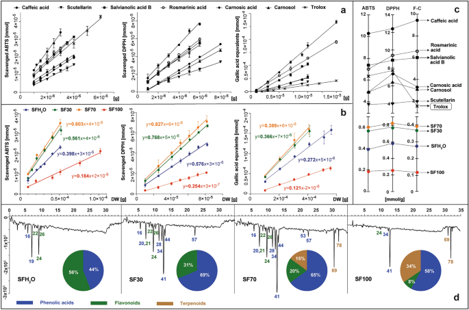
The presented study compared the results of the three most popular spectrophotometric tests using ABTS, DPPH and Folin–Ciocalteu (F–C) reagents. ABTS and DPPH assays are used widely to determine free radical scavenging activity of extracts, as are pure compounds. For S. fruticosa extracts, the calculated antioxidant activity describes the number of ABTS or DPPH molecules reduced by antioxidants derived from 1 g of dried material after 10 min of reaction. These values were calculated in the linear range of the method and expressed as the slope of the line describing the relationship between the number of reduced millimoles of oxidants and various amounts of tested samples – as grams of dry matter in reaction mixtures (Fig. 2b).
The antioxidant activity of standards (caffeic acid, scutellarin, salvianolic acid B, rosmarinic acid, carnosic acid, carnosol and trolox) and S. fruticosa extracts: SFH2O–water extract; SF30–30% ethanol extract; SF70–70% ethanol extract; SF100–ethanol extract, tested in vitro with ABTS, DPPH and F–C reagents presented as plots showing the dependency curves of reagent reduced by tested standards (a) or extracts (b) and expressed as slopes of the curves equal to the milomoles of reagent reduced by 1 g of tested sample (c) set with the antioxidant profiles of extracts, registered at 734 nm after post-column derivatization with ABTS, with the main classes of antioxidants on the pie charts (d). For the identity of peaks, see Supplementary Table S1.
The study also includes the method with the Folin–Ciocalteu reagent. It consists in the transfer of electrons in an alkaline environment from compounds with active hydroxyl groups to phosphomolybdic phosphotungstic acid complexes. The reducing capacity in this case was expressed as the number of millimoles of gallic acid equivalents, that formed blue complex and were derived from 1 g of dry matter of the plant. The same approach was used for the selected pure substances present in sage, such as: caffeic acid, carnosic acid, carnosol, salvianolic acid B, scutellarin, rosmarinic acid and additionally for the reference antioxidant—trolox (Fig. 2a). Such a method of determining and calculating the antioxidant activity of plant material and pure substances was described previously by Kusznierewicz et al.27 and Baranowska et al.28, respectively. The resulting slope values were plotted on separate axes for each test conducted for standards and samples (Fig. 2c). Each of the tested phenolic standard exhibited antioxidant activity, increasing inorder as follows: scutellarin < carnosol < carnosic acid < salvianolic acid B < rosmarinic acid < caffeic acid. Three of them – salvianolic acid B, rosmarinic acid and caffeic acid – were more efficient than trolox, a compound used commonly as reference in antioxidant activity determination assays. The antioxidant activity of all studied extracts of S. fruticosa was dose dependent in ABTS assay as well as in the DPPH test. Therefore, as the amount of extract added to the reaction mixture increases, so does the reducing power towards these radicals. The lowest total antioxidant activity was observed for the SF100 extract, followed by almost two times higher for SFH2O, and nearly four times higher for SF30 and SF70. The results of the F–C test followed the same trend as ABTS and DPPH with a Pearson correlation of 0.99, which indicates that the antioxidant activity of extracts depend greatly on the content of phenolics, as demonstrated by Lantzouraki et al.29.
Based on the contents of 6 phytochemicals selected for testing in the extracts (Table 1) and the antioxidant activities determined for them and for the extracts (Fig. 2a,c), we can determine the estimated contribution of these compounds to the total antioxidant activity of individual extracts. In the case of SFH2O, SF30 and SF70 extracts, 6 selected compounds, depending on the test, theoretically covered 21–30%, 45–63% and 64–86% of the determined total antioxidant activity, respectively. These results suggest the possible presence of other additional antioxidants in these extracts and/or their synergistic effects. Only in the case of the SF100 extract did the sum of the activities of the 6 standard compounds exceed the determined total activity of this extract ranging from 20 to 49%, depending on the test used. Such an observation may be the result of possible antagonistic interactions between the phytochemicals present in this kind of extract.
More detailed information on the types of antioxidants present in the tested S. fruticosa extracts was provided by using HPLC post-column derivatization with the ABTS reagent. The antioxidant profiles obtained by this method, as well as the contribution of different classes of antioxidants in the total antioxidant activity, are shown in Fig. 2d. In addition to the 6 standard antioxidants tested earlier, the S. fruticosa extracts also contained other antioxidants such as przewalskinic acid A, salviaflaside, luteolin rutinoside, luteolin glucoside, isorhamnetin glucuronide, coumaroyl caffeoylglycoside, salvianolic acid K and salvianolic acid F. Profiles characterized by the largest number and size of negative peaks indicating the reduction and discoloration of ABTS radicals were observed for the SF70 and SF30 extracts. Despite the similarity of the antioxidant profiles of these two samples the intensity of common signals was higher for SF70 and additional activity originating from diterpenoids was also noticed. Only in the profiles of SF70 and SF100 extracts were negative peaks originating from diterpenoids observed, with their share in the total antiradical activity at 15 and 34%, respectively. The main antioxidant in all the extracts containing ethanol was rosmarinic acid – the most abundant phenolic acid and one of the strongest antioxidants among studied standards. The same result was also reported for S. officinalis and S. hispanica extracts30,31.
In aqueous extract (SFH2O) the antioxidant activity originated mainly from two compounds: przewalskinic acid A and scutellarin, as rosmarinic acid extraction with water alone was less effective.
Xanthine oxidase inhibitory activity
The enzyme xanthine oxidase (XO) catalyzes the oxidation of hypoxanthine and xanthine to uric acid, an excess of which in the blood causes gout to develop. During XO reoxidation, molecular oxygen acts as an electron acceptor, producing a superoxide radical and hydrogen peroxide. Consequently, XO is considered an important biological source of superoxide radicals which, together with other reactive oxygen species, contribute to the body’s oxidative stress and is involved in many pathological processes such as inflammation, atherosclerosis, cancer, ageing, etc.32. A recent therapeutic approach to hyperuricemia treatment is to inhibit the XO enzyme. Various drugs containing XO inhibitors (allopurinol, febuxostat) have been developed, the use of which is unfortunately associated with certain side effects. For this reason, there is a constant search for natural XO inhibitors that could provide an alternative to these synthetic compounds. There are some reports in the literature about the ability of several species of Salvia (S. plebeia, S. miltiorrhiza, S. verbenaca) to inhibit XO33,34,35, therefore, the possible occurrence of this activity was also tested in the studied S. fruticosa extracts. In addition, XO inhibitory activity was also determinedin selected phenolic compounds typical for sage such as: caffeic acid, carnosic acid, carnosol, salvianolic acid B, scutellarin, rosmarinic acid and additionally, for reference, the XO inhibitor allopurinol.
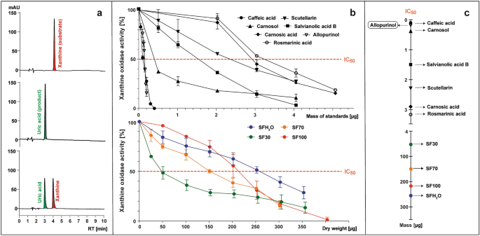
The transformation of xanthine (substrate) to uric acid (product) by XO with or without the presence of tested samples was monitored with the use of HPLC-PAD at 285 nm (Fig. 3a). The enzyme activity was calculated as the percentage of the uric acid peak area formed in the presence of the tested sample compared to the control without the addition of the sample (Fig. 3b). The inhibition of the XO enzyme was expressed as an IC50 value, meaning the mass of standard or dry weight of sample (μg) capable of reducing enzyme activity to 50% (Fig. 3b,c).
The examples of HPLC chromatograms at 285 nm of post-reaction mixtures containing (from top): xanthine; xanthine and xanthine oxidase (XO); xanthine, XO and inhibitor (a), which were the basis for preparing the plots representing the curves of XO activity in the presence of tested standards (caffeic acid, scutellarin, salvianolic acid B, rosmarinic acid, carnosic acid, carnosol and allopurinol) or S. fruticosa extracts (SFH2O–water extract; SF30–30% ethanol extract; SF70–70% ethanol extract; SF100 – ethanol extract) (b), which were used to determine the parameter IC50, meaning the micrograms of tested sample needed to reduce XO activity to 50% (c).
The known XO inhibitor allopurinol was used as a reference, with an IC50 value of 0.15 μg (5.5 µM). All the studied standards showed the XO inhibitory activity with an IC50 ranging from 0.1 to 3.15 μg (2.8–43.8 µM). XO inhibitory activity increased in order as follows: rosmarinic acid < carnosic acid < scutellarin < salvianolic acid B < carnosol < caffeic acid. Caffeic acid showed the lowest IC50 value (0.1 μg; 2.8 µM), indicating the strongest XO inhibitory activity among the tested compounds. It was even stronger than allopurinol, which is inconsistent with the data presented by Wan et al.36 and Flemmig et al.37, where the IC50 of caffeic acid was almost 8 or 2 times lower than that of allopurinol, respectively. These differences may result from the origin of the XO selected for the tests. In the cited studies, an oxidase from bovine milk was used, while for this study a microbial oxidase was selected. In this study, rosmarinic acid had the lowest to inhibit XO (3.2 μg; 43.8 µM), but Ghallab et al.38 reported that synergistic combination of allopurinol and rosmarinic acid can lower the dosage of synthetic drugs needed. XO was inhibited by all studied S. fruticosa extracts, although over 1000 times less effectively than by allopurinol, in line with data reported for other Salvia species. Scutellarin and other flavones have been described previously as strong inhibitors of XO39. Despite only a slight difference of total flavonoids content between SF30 and SF70 extract, and the contents of other anti-inflammatory compounds being more favorable for the SF70 extract, SF30 had the strongest ability to inhibit the XO activity. The IC50 value for SF30 was 50 μg and based on this parameter, the potential anti-inflammatory activity of SF70, SF100 and SFH2O extracts was determined as 3, 4 and 5 times weaker, respectively.


